
Atresia is a defect on the calf that does not allow them to adequately take in or excrete food because of a blockage or narrowing in their digestive system. The defect can be as far up as the esphagus, or as far back and the anus.
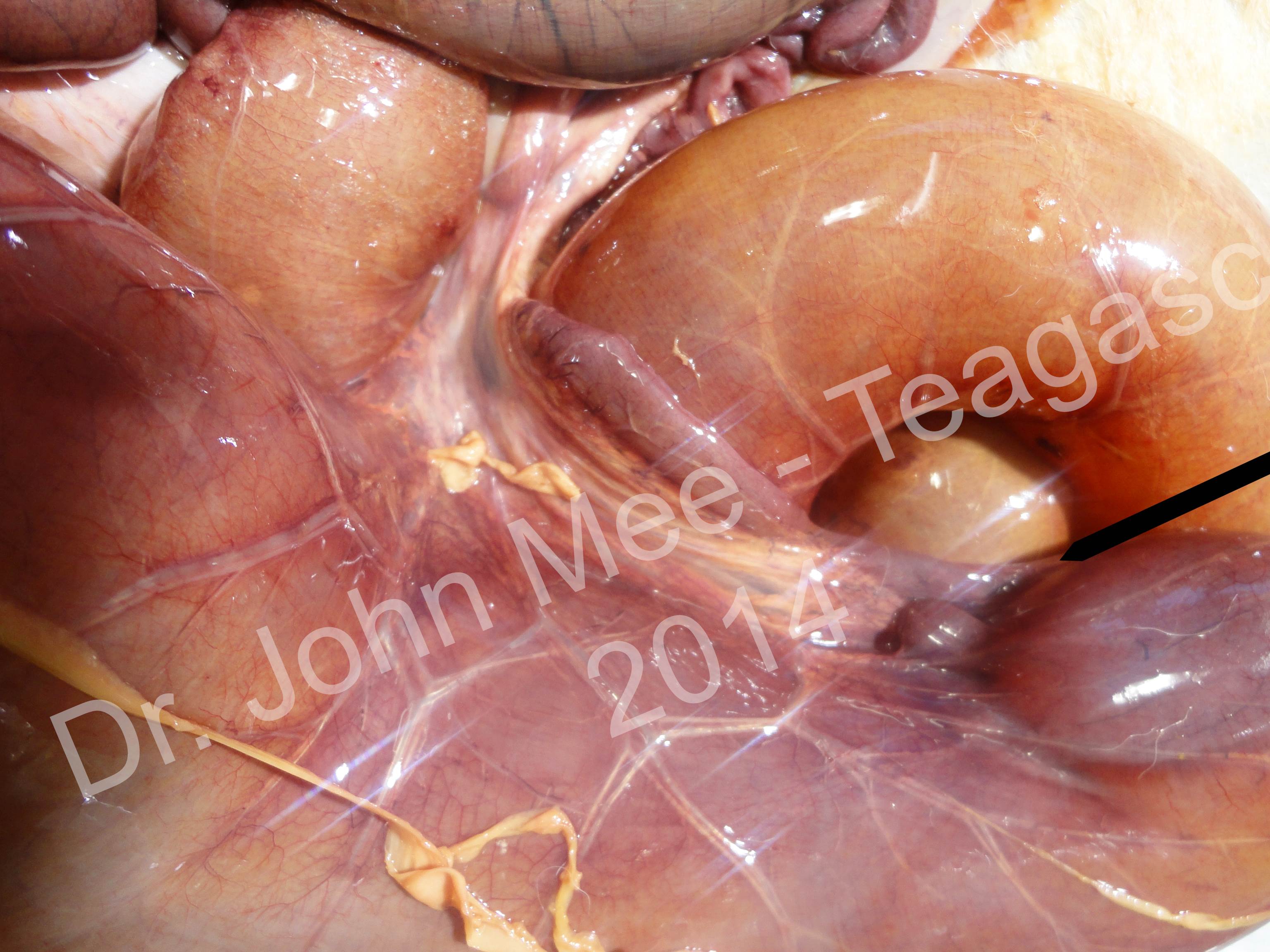
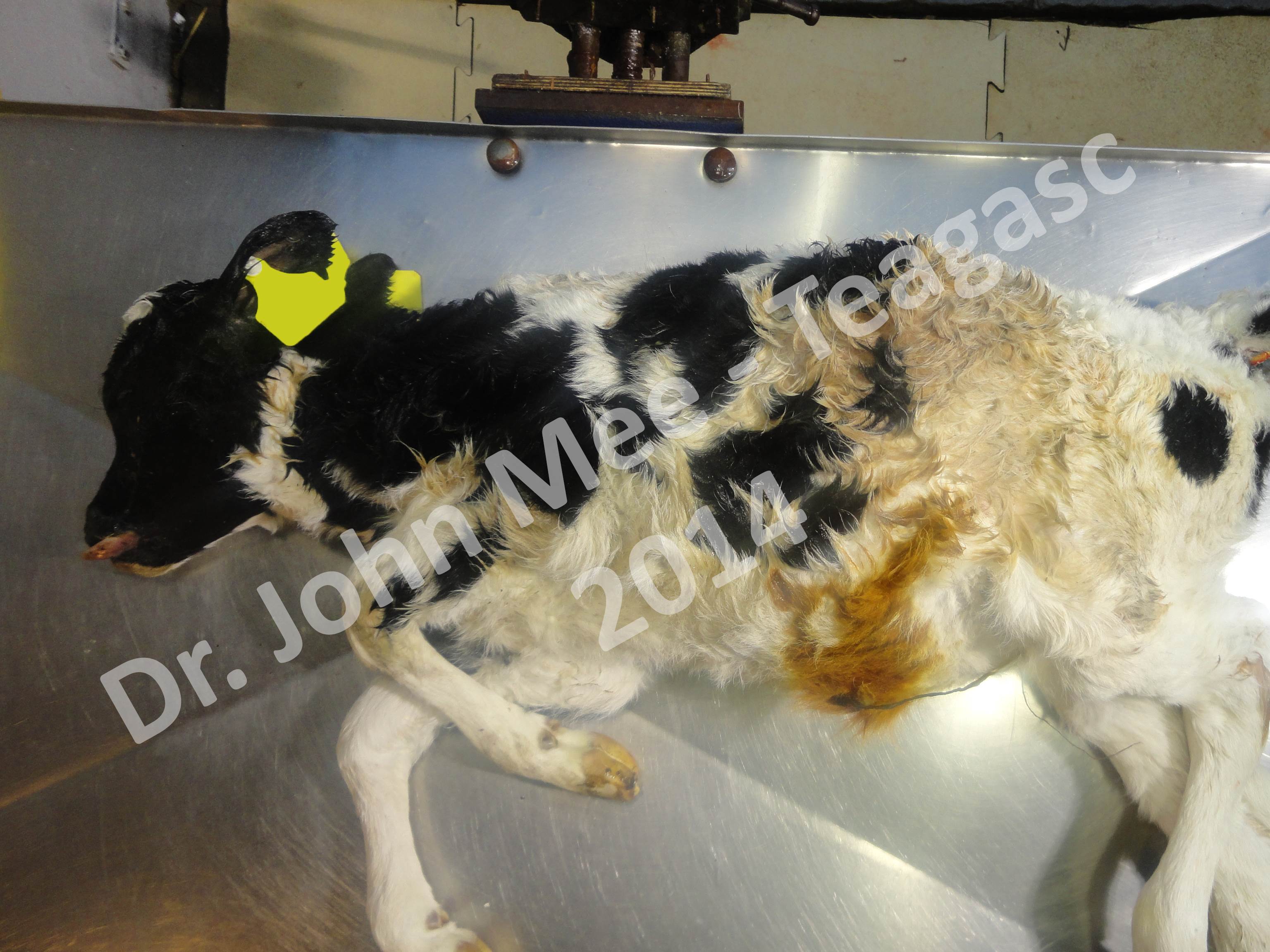
Atrasia ani has been described as an animal “Without a back passage,” meaning they do not have an anus. Atresia jejuni and Atresia coli occur in the intestine of the animal, specifically as the names suggest, the jejunum and the colon. Most of the time these calves are born healthy and do not have any visible defects. It is only after they have suckled that a problem occurs because they cannot pass feces. This will result in a paunched gut and the animal becoming very sick quickly. Surgery can be performed on these animals, but it is often better to have them euthanized because an animal may have multiple spots where these blockage occur.
If the animal has a post-mortem performed, it would be of interest to us if the animal had multiple areas of atresia and where they were found in the animal because right now it is unknown it each type of atresia has a different mutation, or if it is a single mutation in the genes that cause the problem.